
Top Essay Writers
Our top essay writers are handpicked for their degree qualification, talent and freelance know-how. Each one brings deep expertise in their chosen subjects and a solid track record in academic writing.
Simply fill out the order form with your paper’s instructions in a few easy steps. This quick process ensures you’ll be matched with an expert writer who
Can meet your papers' specific grading rubric needs. Find the best write my essay assistance for your assignments- Affordable, plagiarism-free, and on time!
Posted: August 20th, 2023
The availability of dental bonding agents for a wider range of clinical application in recent year has emphasized the need for a reliable ad convenient methods of evaluating adhesive performance. The shear bond strength test method has been criticized due to variation among the results of various studies and also its validity under clinical condition [50] [51]. Yet the test is highly popular even today, due to its simplicity [52]. This model tests the shear strength between two materials that are connected via adhesive agent. The load is applied till the point of fracture. The bond strength is calculated by dividing the applied force required to disassociate the assembly by the cross-sectional surface area of the bonded dentin.
The current generation of dentin bonding agents often exhibit cohesive type of failure [53] [54]. It may have led to the belief that dentin bonding agents have acquired enough strength, that there is no need for further improvement [55]. But it is a well-known fact that mechanically properties of resin is inferior to dentin, hence it questions the cohesive sequelae of dentin failure [52]. Previously, many theories were proposed to explain the reasons for cohesive dentin failure in the area of bonding, but it was Gwinnett who suggested that dentin is weakened by dehydration and the substrate involved is the hybrid layer [29]. Infact, it’s a proven fact that failure at the interface is non-homogenous [56]. Therefore, there are multiple factors like not only the mechanical properties but also alteration in the shear mechanics that can lead to monolithic cohesive type fracture in dentin [52].
We get a lot of “Can you do MLA or APA?”—and yes, we can! Our writers ace every style—APA, MLA, Turabian, you name it. Tell us your preference, and we’ll format it flawlessly.
Clinical Research Associates (CRA) newsletter report from July 2002 [57] indicates some characteristics of an ideal shear bond test method. In this report, usage of human dentin is encouraged. Failure usually occurs within the adhesive layer, at the adhesive-tooth junction and / or at the adhesive restorative junction. Many methods have been developed to test the adhesive for bond strength out of which single plane lap test is the most common.
The CRA newsletter report indicates that the higher the percent of samples that have the three modes of failure shown in figure 26, the better the test method is. The shear bond tester used in this study is Ultratester© (UltraDent, UT, USA), which has been commonly used for shear bond testing in dental research.
Multiple Infrared spectroscopy(IR spectroscopy) is the study of a part of the electromagnetic spectrum, mainly in the infrared region. The infrared spectrum has longer wavelength that visible light. The main utility of the infrared spectroscopy is that it can be used to identify and evaluate different chemicals. The most common instrument used is Multiple Attenuated Internal Reflection – Infrared Spectroscopy. When an infrared beam is passed through the sample, it produces a unique spectrum and a vibrational frequency which is where the absorption is recorded in this technique.
Starts at $10/page for undergrad, up to $21 for pro-level. Deadlines (3 hours to 14 days) and add-ons like VIP support adjust the cost. Discounts kick in at $500+—save more with big orders!
MAIR-IR is a very sensitive surface characterization technique that allows the operator to visualize materials containing functional groups or covalent bonds on a prism that can internally reflect an emitted IR beam [72]. Covalent bonding is necessary because there are no bond vibrations for ionic bonds. Additionally, it is important to note that this technique offers no data on the physical form of the sample, only the bonding chemistries.
The multiple reflection process displays more sensitive characteristics, compared to its predecessor which worked on the principle of single reflection. This form of IRS requires an intimate contact between a sample and the prism used to create the multiple reflections. The prisms in question are made of Germanium and Thallium Bromoiodide (KRS-5), two materials that exhibit favorable indices of refraction and transparency in the infrared range (~2-20μm). Germanium, mined primarily from sphalerite, was used when examining thin or liquid films, while the KRS-5 elements were utilized when analyzing samples with greater thickness. As shown in Fig. 7, infrared light is broadcast through the prism, entering at a 90° angle.
Following its entrance into the prism, the infrared beam is reflected every time that the angle of incidence is greater than the critical angle. At the point of each reflection, an evanescent wave is created. When these evanescent waves are triggered there is no loss in beam energy unless the wave is able to resonate with anything making an intimate contact with the prism. The depth of penetration, calculated using formula (1), into the sample is a function of the beam wavelength, incident angle, and the refractive indices of the reflector and sample [1, 2].
100%! We encrypt everything—your details stay secret. Papers are custom, original, and yours alone, so no one will ever know you used us.
 (1)
(1)
The subsequent beam that is transmitted out of the prism possesses less energy than when it entered due to these reactions. This energy loss is attributed to the absorption of the infrared light by varying molecules existing within the sample surface. By comparing the amount of reflected light to the infrared frequencies, a valid spectrum is created. The molecules present within the sample display different amount of energy absorption based on their bonds. It is because of this that the peaks can be used to indicate the varying types of molecules within the sample. By comparing these sample compositions to known spectra or known material compositions, unknown materials can be identified with relative certainty.
The procedure for IRS analysis remains same with Germanium prism as well as with KRS-5. Germanium is a very expensive, relatively brittle material, with a polished, spectral surface. For these reasons, germanium is a great prism to use if seeking the chemistry of a liquid or a sample dissolved in a liquid. Other uses include characterization of adhesives, and when using a horizontal unit so that the prism may lay flat, the MAIR-IR can take multiple analyses during a drying period for time-dependent comparison. KRS-5 prism is conducive for solid samples. It can be still used liquid samples, but the fact that KRS crystals are water soluble must be taken into effect. Water can affect the surface properties of a KRS prism, thus making results from IRS inaccurate. To avoid the chance of this occurring, thick, non-liquid samples are often used. Clamping is involved on one or both sides of the prism to create an intimate contact.
The stereo or dissecting microscope is a variant of optical microscope for low magnification observation of a given specimen. It utilizes the light reflected from the specimen, rather than the light transmitted through it to obtain an image. The microscope has two separate optical paths with two objectives and eyepieces, in order to enable different viewing angles. It is this feature that facilitates, a three-dimensional visualization of the sample being examined. For recording and analyzing samples with complex surface topography, wherein a three-dimensional view is required for examination, stereomicroscope overhauls other techniques.
Nope—all human, all the time. Our writers are pros with real degrees, crafting unique papers with expertise AI can’t replicate, checked for originality.
The main two types of magnification systems in stereo microscopes are fixed and zoom. The fixed magnification as the name suggests, is achieved by setting a fixed degree of magnification. Zoom/pancreatic type of magnification allows a continuous range and degree of magnification depending on the topography. It also enhances the total power of magnification by a set factor through use of auxiliary objectives [56].
A video charged coupled device (CCD) camera is available to capture images of the specimens. These can be fitted within the microscope and the image obtained from both the eye piece is converted by special software, in order to integrate and produce a 3-dimensional image. The results are viewable through the use of 3D Glasses. However, in spite of availability of these dynamic camera technology, generally a camera attached to just a single eyepiece is used to record conventional 2D images.
In scanning electron microscopy (SEM), a beam of electron is subjected across the sample surface. The signals generated in response to the collision are recorded. The receptor detects specific signals, required to generate a sample elemental composition. The signals mostly accounting for greatest amount of information in SEM are the secondary electrons, backscattered electrons and X-rays [57].
Our writers are degree-holding pros who tackle any topic with skill. We ensure quality with top tools and offer revisions—perfect papers, even under pressure.
Emission of secondary electrons from the surface at the top, will produce an interpretable image of the surface. Whereas, the primary electron is responsible for the image contrast due to its small diameter [57]. The reflected primary electrons are called as backscattered electrons. Unfortunately, the image contrast of these samples are not as fine as those of secondary electrons. The contrast of an image depends on the atomic number of the sample, whereas the back scatter electrons are emitted from a depth in the sample compared to the secondary electrons [57].
Another essential aspect about understanding the theory behind surface energy has further reasoning embedded in the theory of thermodynamics and a simple understanding of the phases of matter. It has been postulated that, “the surface tension of a liquid is exactly equal to its thermodynamic surface free energy”. For a solid, however, this concept is not necessarily congruent. The reason for this is that a solid cannot achieve equilibrium. A liquid is constantly in flux, or is considered to be fluid. The molecules within are not limited to one arrangement, but are instead constantly moving and rearranging. The molecules in a solid are static, and have been isolated to a single configuration since the moment it entered the solid state. Because of this, there may be molecular variations at different locations on the surface of a solid material. What this means for contact angle measurements is that a prudent researcher should be taking multiple measurements at different locations on the material to correct for the inherent error in working with these non-equilibrium solids. While duplicates in a scientific protocol are standard procedure, the requirement for it in contact angle measurements is absolutely essential. As a result, the standard error when using a goniometer is ±2°.
Experts with degrees—many rocking Master’s or higher—who’ve crushed our rigorous tests in their fields and academic writing. They’re student-savvy pros, ready to nail your essay with precision, blending teamwork with you to match your vision perfectly. Whether it’s a tricky topic or a tight deadline, they’ve got the skills to make it shine.
Zisman plot [61]
The surface tension at a solid/gas interface can be infered from a Zisman Plot. This measurement of variations in contact angles acts as the function of the known surface tension,γLV, for a series of liquids. The experimentally obtained dependence ofcos(θ)onγLVis often linear. The value for whichcos(θ)extrapolates to 1 is termed the surface tension,γSV, of the solid/gas interface. An example of a typical Zisman Plot is shown in Figure 29.
The rationale is as follows:
Guaranteed—100%! We write every piece from scratch—no AI, no copying—just fresh, well-researched work with proper citations, crafted by real experts. You can grab a plagiarism report to see it’s 95%+ original, giving you total peace of mind it’s one-of-a-kind and ready to impress.
a liquid with a surface tension equal or slightly less than the surface tension of a particular material will wet that surface, i.e. the contact angle will be 0o. However, this widely-used approach can be applied only under proper conditions. The Young equation atθ = 0gives: γSV=γSL+γLv.
But notγSV= γLVas obtained from the Zisman approach.
The liquid and solid studied must have an interface withγSL≈0. This is approximated in the case of organic polymer solids and liquid saturated hydrocarbons or aqueous solutions of saturated alcohols at sufficiently high concentrations (>20%). This is the reason why the surface tension obtained from a Zisman plot is often referred to in the literature as the critical surface tension of wetting but not the surface tension of a solid/gas interface.
Method
Direct Measurement (Telescope-Goniometer)
The practice of direct measurement of the tangent angle at the intersection of solid-liquid-gas-phase contact point on a stalkless drop profile is the most common for obtaining a contact angle measurement. Bigelow and co-workers set up a simple and convenient instrument, which they referred to as a “telescope-goniometer” to measure contact angles of various liquids on polished surfaces [62]. In 1960s, W.A. Zisman designed the first contact angle goniometer for commercial use, was manufactured by ramé-hart corporation (Fig. 31). Contact angle analysis requires the use of a reasonably simplistic setup. A green light is used to illuminate the outline of a drop that has been placed on a test material. The contact angle, θ, is measured by means of an optical goniometer which magnifies the drop 10 times its original size. In the early history of contact angle measurement, Zisman and his co-workers used platinum wire [63] to form sessile drops of liquids on solid substrates. A fine platinum wire (about 8-10 cm long and as thin as 0.05–0.10 mm in diameter) was first cleaned using the Bunsen burner by heating to red. As soon as the red color resolves back to normal color the wire is dipped into the liquid and gently flicked. This flicking action leads to formation of a pendant like droplet that hangs from the wire-tip. The drop forms a sessile drop when brought in contact with the solid surface. Spelt et al [64] claimed a reproducibility of +/- 2°, but certain concerns regarding the kinetic energy related to the flow of the droplet and the uncontrolled deformation occurring while removal of the platinum wire, leading to formation metastable contact angles.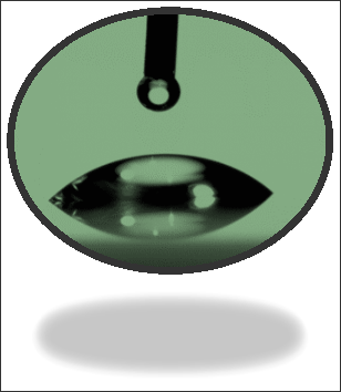

Using the adjustable rings around the ocular piece, an operator can determine the approximate contact angle that the liquid makes with the surface. With the adjustable ring, the operator must form a line tangent to the liquid droplet and the corresponding angle measurement defines the angle through the liquid (Fig 32). After taking this measurement, the process should be repeated on the opposite side of the liquid. Following this data point, another drop of liquid must be placed upon the existing droplet. Again, the operator must measure both sides of the liquid against the material surface. After repeating this process once more, the researcher has one full data point for a test liquid of a known surface tension. Typically this first liquid is water, and the following liquids can be referenced in Appendix A.
Once the operator has collected many data points (11 liquids were used in this protocol), these can now be plotted on the aforementioned Zisman Plot. Instead of 6 data points for each liquid, an average is made and the cosine of that average angle is on the y-axis of the plot. Along the horizontal axis is the pre-defined surface tension of the test liquid. The unit used is dynes per centimeter which defines the amount of force per unit of length. The liquids are pure samples that are nearly perfected with great care to achieve optimal results. The student set used in this protocol are of slightly different surface tensions than the Master Set used in most circumstances. Once the data has been fully input into the plot, the operator must create a best-fit line that traverses the upper limit of the graph
(or the cos=1) . The point at which the best-fit line crosses is, as previously mentioned, the CST.
Following data collection and plotting it on Zisman’s Plot, an adjunct approach is to take the aid of a custom-made software developed in University at Buffalo for contact angle analysis. The objective analysis and interpretation of the data gives three main results i.e. critical surface tension, gamma-P (polar forces) and gamma-D (dispersion forces) portions. The results obtained for gamma-P (polar forces) and gamma-D (dispersion forces) are vital. Combined, as stated in the introduction, these equate to the γSV, or the surface free energy. Also the program gives a value of the best estimate of the CST with the data provided. This value may or may not represent the true CST of the solid and this is where the obscuring polar forces appear. If the polar liquids interact with the material and yield inaccurate data points, and the program does not automatically exclude this data, the results will suffer at this location. In case the operator suspects any discrepancy or poor data collection of certain values, due to a liquid’s interaction with the material, those data can be excluded without having any impact on the final result.
Tags: Academic Paper Assistance, Affordable College Homework, APA Citation Assignment Help, Assignment Help AustraliaYou Want The Best Grades and That’s What We Deliver
Our top essay writers are handpicked for their degree qualification, talent and freelance know-how. Each one brings deep expertise in their chosen subjects and a solid track record in academic writing.
We offer the lowest possible pricing for each research paper while still providing the best writers;no compromise on quality. Our costs are fair and reasonable to college students compared to other custom writing services.
You’ll never get a paper from us with plagiarism or that robotic AI feel. We carefully research, write, cite and check every final draft before sending it your way.